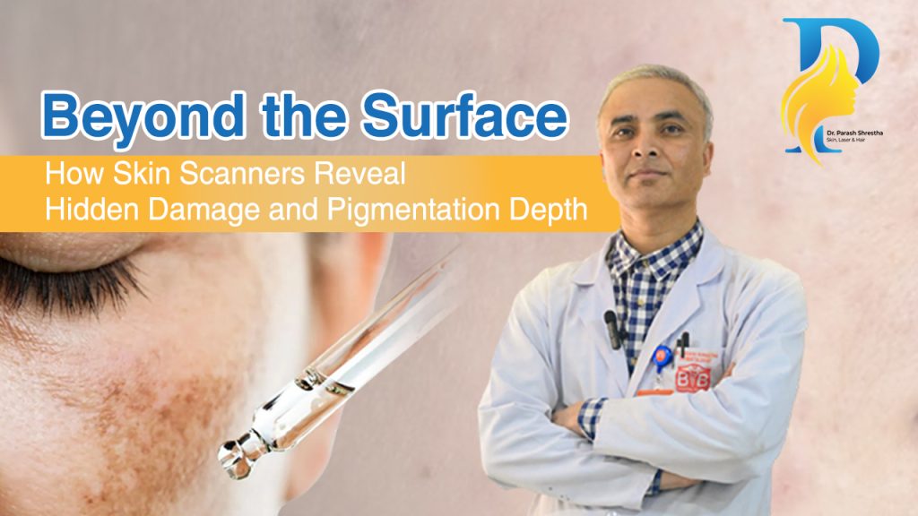
Beyond the Surface: How Skin Scanners Reveal Hidden Damage and Pigmentation Depth
When you look in the mirror, you only see the “final result” of your skin’s health: the visible spots, the fine lines, and the uneven tone. However, the human eye is remarkably limited. It cannot see the damage brewing in the deeper layers of the dermis, nor can it accurately measure the depth of pigmentation that hasn’t surfaced yet. For years, patients would visit a Dermatologist in Kathmandu or a Dermatologist in Lalitpur describing “sudden” dark spots. In reality, these spots weren’t sudden; they were years in the making. Today, advanced skin analysis machines have revolutionized how a doctor diagnoses and treats these issues, moving from subjective observation to objective, data-driven science. The Anatomy of Pigmentation: Why Depth Matters To understand how a scanner works, we must first understand the structure of the skin. Your skin is not a single sheet; it is a complex, multi-layered organ. 1. The Epidermal Layer (The Surface) This is the outermost layer. Pigmentation here is often “new” or the result of recent sun exposure. Spots in this layer are usually easier to treat with topical creams or light chemical peels because the cells turn over every 28–40 days. 2. The Dermal Layer (The Deep Tissue) The dermis sits beneath the epidermis. When pigmentation reaches this depth often due to chronic sun damage, hormonal changes (Melasma), or deep inflammation it becomes “entrenched.” Surface-level treatments cannot reach this depth effectively. 3. The Dermal-Epidermal Junction (DEJ) This is the “basement membrane” where the two layers meet. Damage here often requires sophisticated laser intervention. How Do Skin Scanners See the “Invisible”? Advanced diagnostic tools, like those used by a Dermatologist in Nepal, utilize three primary types of imaging technology to map your skin’s health. RGB (Visible Light) Imaging This captures what the human eye sees but at a much higher resolution. It documents the current state of your skin, including visible pores, redness, and surface-level spots. It serves as the “baseline” for your medical record. Cross-Polarized Lighting This technology eliminates the “glare” and reflections from the skin’s surface. By doing so, it allows the doctor to see the vascular (blood vessel) structure and underlying inflammation. This is crucial for diagnosing conditions like Rosacea or hidden irritation that might be causing “phantom” sensitivity. UV (Ultraviolet) Fluorescence This is the “time machine” of dermatology. UV light causes certain substances in the skin, such as porphyrins (bacteria) and melanin (pigment), to glow or darken. It reveals sun damage that is currently hidden beneath the surface but is guaranteed to emerge as a dark spot in 1–3 years if left untreated. Technology What it Detects Clinical Utility RGB Imaging Visible spots, wrinkles, pores Progress tracking & baseline Polarized Light Redness, inflammation, thin skin Sensitivity & vascular diagnosis UV Imaging Subsurface melanin, sun damage Prevention & deep-pigment mapping Why All Spots are Not Created Equal A common frustration for patients is why a “whitening cream” works for a friend but not for them. The answer lies in the pigmentation depth. Without a scan, even an experienced Dermatologist in Kathmandu might find it challenging to distinguish between epidermal and dermal melasma through visual inspection alone. The Role of the Skin Analysis Machine A professional scanner provides a “Skin Score.” This score compares your skin against a global database of thousands of individuals of the same age and skin type. Top 5 Benefits of Professional Skin Analysis in 2026 1. Early Detection of Sun Damage Skin scanners can detect “latent” pigment. If you see a cluster of dark spots on a UV scan that aren’t visible in the mirror yet, you can start using specific antioxidants and higher-grade SPF to prevent them from ever surfacing. 2. Accurate Treatment Selection Should you get a Carbon Peel, a chemical peel, or a Q-Switched Laser? The scanner decides. Epidermal spots respond well to peels, but dermal pigmentation usually requires the specific wavelength of a laser to shatter the deep-seated melanin. 3. Monitoring Treatment Efficacy Often, deep treatments take months to show surface results. A scanner allows your Dermatologist in Lalitpur to show you that the pigment is breaking up underneath, even if you don’t see it yet. This keeps patients motivated and compliant. 4. Personalized Product Matching In 2026, “one size fits all” skincare is dead. By analyzing pore size and sebum (oil) levels, the machine helps the Doctor recommend products that won’t clog your pores or dry out your barrier. 5. Identifying Biological vs. Chronological Age Many modern machines now offer a “TruAge” feature. This tells you if your skin is behaving like a 30-year-old’s or a 50-year-old’s based on elasticity and damage levels. The 2026 Landscape: Dermatology in Nepal Nepal has seen a significant shift in medical aesthetics. Finding a qualified Dermatologist in Nepal is now easier, but choosing the right clinic depends on the technology they utilize. At the clinic of Dr. Parash Shrestha, the focus is on “Integrative Diagnostics” combining clinical expertise with high-tech imaging. Whether you are looking for a Dermatologist in Kathmandu for acne or a Dermatologist in Lalitpur for aging concerns, always ask: “Do you perform a subsurface skin analysis before starting treatment?” Case Study: The “Invisible” Melasma Consider a patient who complained of a small patch of darkness on their cheek. To the naked eye, it looked like a simple sunspot. Upon scanning with a multispectral device, the doctor discovered that the pigment was actually part of a much larger, symmetrical “mask” of Melasma that was deeply rooted in the dermis. Instead of a standard exfoliating facial, the patient was started on a specialized medical protocol. Without the scan, the patient would have wasted time and money on treatments that couldn’t reach the required depth. Understanding the Results: What Your Report Means When you receive your skin analysis report, you will likely see several categories: Frequently Asked Questions (FAQ) Is a skin scan painful? No. It is a completely non-invasive procedure. You simply place your face in a specialized booth or cradle, and several high-resolution photos are taken with
Beyond the Surface: How Skin Scanners Reveal Hidden Damage and Pigmentation Depth Read More »